7 min read
Hip Osteonecrosis: Signs and Treatment Options
Early signs of hip osteonecrosis, why MRI can matter and when joint-preserving care or replacement is considered.
Hip Osteonecrosis: Signs and Treatment Options
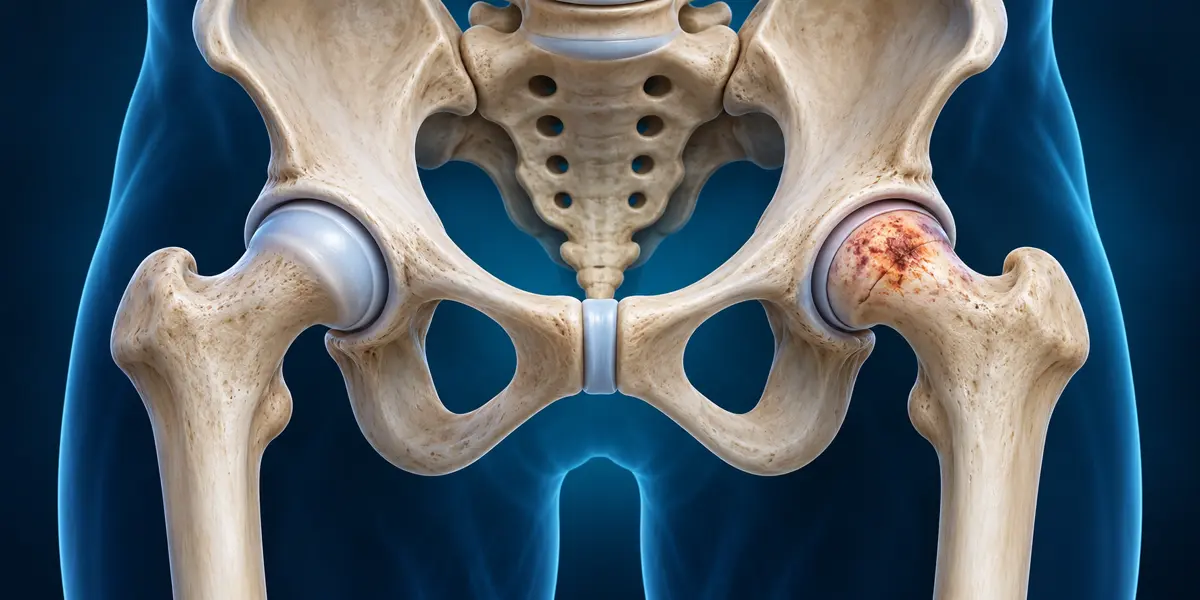
Hip pain is often blamed on a muscle strain or ordinary arthritis. In some people, however, the problem is osteonecrosis, also called avascular necrosis. This means part of the bone has been damaged after its blood supply is reduced. In the hip, the rounded head of the thigh bone is the area most often discussed. Recognising the condition early matters because treatment choices change when the joint surface begins to collapse.
This article explains what hip osteonecrosis can feel like, which tests may clarify it, and why hip replacement is not the automatic first answer. It cannot tell you the stage of your own hip from symptoms alone. If pain is persistent or worsening, an orthopedic assessment can help identify the cause and protect function.
What happens inside the hip?
The femoral head is the ball that moves within the hip socket. Bone is living tissue and depends on a blood supply. When circulation to an area is interrupted, the affected bone may weaken. Over time, the smooth joint surface can lose its shape or collapse, contributing to pain and arthritis. The timing and severity vary; the condition is not identical in every patient.
Osteonecrosis can follow an injury such as a hip dislocation or fracture. Non-traumatic cases may be associated with factors such as long-term or high-dose corticosteroid exposure, heavy alcohol use, or certain medical conditions. Some people have no obvious cause. A clinician should review the history without assuming that one risk factor proves the diagnosis.
The US National Institute of Arthritis and Musculoskeletal and Skin Diseases notes that early disease may cause no symptoms. Later, pain is commonly felt in the groin, and sometimes in the buttock. It may first occur with weight bearing and eventually appear at rest. These symptoms overlap with other hip problems, so imaging and examination are important.
Early warning signs worth discussing
The most useful clue is not one dramatic symptom. It is a pattern: deep groin or hip pain that returns, worsens while walking or standing, and begins to limit movement. A person may limp or find it harder to put on shoes, climb stairs, or rotate the hip. Pain can also be felt around the thigh or knee, which is why the true source may be missed.
Seek an assessment sooner if the pain follows a hip injury, if you have relevant risk factors, or if symptoms are steadily increasing. Sudden severe pain or inability to bear weight needs urgent evaluation because it could reflect a fracture or another acute problem, not only osteonecrosis.
Our guide to hip pain while walking reviews several possible causes. This article focuses narrowly on what changes when osteonecrosis is suspected or confirmed, so the two pieces answer different questions.
Can an X-ray miss early osteonecrosis?
Yes. A plain X-ray is often a useful first imaging study because it can show changes in the bone and joint surface, especially later in the disease. But an early X-ray can look unremarkable even when symptoms and risk factors raise concern. According to the NIAMS diagnostic guide, MRI is highly sensitive and can detect early changes before they appear on an X-ray.
That does not mean everyone with hip pain needs an MRI. The decision depends on examination, the X-ray, history, and whether a more sensitive test would change management. MRI can also help an orthopedic specialist estimate the amount and location of affected bone. Those features, along with whether the joint surface has collapsed, matter more for treatment planning than the word "necrosis" alone.
If you already have an MRI report, bring the images as well as the written interpretation. A specialist may need to compare imaging with your symptoms and examination rather than rely on one line of the report.
What treatment is possible before collapse?
Early-stage care may include reducing stress on the joint, pain management, and addressing relevant risk factors. A cane or crutches may help some patients protect a painful weight-bearing hip. Physiotherapy may support movement and function, although it cannot be assumed to restore a damaged blood supply. The treatment plan is individual; do not begin weight-bearing restrictions or exercises from a generic online schedule.
For selected patients before collapse, a surgeon may discuss joint-preserving procedures such as core decompression. This involves creating a channel in the affected bone to reduce pressure and, in some approaches, combining it with other techniques. Its suitability depends on stage, lesion size, location, age, and the surgeon's assessment. No procedure guarantees that the disease will stop.
NIAMS explains that non-surgical care can ease symptoms but often does not cure or reliably stop progression. This is why follow-up matters. A patient feeling a little better after rest should not assume the bone has recovered without the agreed clinical review.
When does hip replacement enter the conversation?
Hip replacement becomes more relevant when the femoral head has collapsed, arthritis is established, pain is substantial, or joint-preserving options are unlikely to work. The operation replaces the damaged joint surfaces rather than treating a small early lesion. It can be an effective option for the right patient, but the decision depends on function, imaging, other health conditions, and individual priorities.
An osteonecrosis diagnosis does not mean "replacement immediately." Equally, delaying assessment until the hip is severely damaged may narrow the available options. Our article on when hip replacement is really necessary discusses the broader decision. The hip replacement recovery guide can help you understand the rehabilitation commitment if surgery is recommended.
For younger or more active patients, timing and implant-related planning are especially important. Ask the surgeon what stage the disease has reached, which alternatives were considered, and what outcome is realistic. A second opinion may be reasonable if the imaging and proposed plan are unclear.
Questions to bring to an orthopedic visit
Ask which part of the femoral head is affected and whether the surface has collapsed. Ask whether an MRI is needed if only an X-ray has been done. Review possible risk factors, including medicines, but do not stop a prescribed steroid or another important treatment without speaking to the prescriber. Ask what activities are safe now, which symptoms warrant urgent review, and how progress will be monitored.
It is also useful to ask what the goal of each option is. Pain relief, preserving your own joint, maintaining walking ability, and reducing future surgery risk are related but not identical goals. A clear plan identifies which outcome is being pursued and what evidence would show whether it is working.
The takeaway
Hip osteonecrosis may begin quietly, and an early X-ray may not show it. Persistent groin or hip pain, especially with relevant risk factors, deserves assessment. MRI can clarify early disease when appropriate, and treatment choices depend heavily on stage and extent of damage. Hip replacement is one option for advanced disease, not an automatic consequence of the diagnosis. Seek qualified orthopedic advice for a plan based on your actual hip rather than a general treatment chart.
Frequently asked questions
Is avascular necrosis the same as osteonecrosis?
The terms are commonly used for the same process: bone damage related to reduced blood supply. The important next question is where it is occurring and how advanced it is.
Can osteonecrosis cause knee pain?
Hip problems can sometimes be felt in the thigh or knee. Knee pain has many other causes too. Examination helps locate the source before deciding which imaging is useful.
Can an MRI diagnose it before an X-ray?
MRI can detect early changes that may not yet appear on a plain X-ray. Whether you need one depends on symptoms, risk factors, examination, and the first imaging results.
Can physiotherapy cure hip osteonecrosis?
Physiotherapy may help mobility and function, but it should not be presented as a cure for damaged bone. A specialist should explain the disease stage and monitoring plan.
Does everyone with hip osteonecrosis need replacement?
No. Some patients may be candidates for monitoring or joint-preserving treatment, especially before collapse. Replacement is more likely when the joint surface is damaged and symptoms significantly limit life.
Should I stop steroids if I have this diagnosis?
Never stop a prescribed steroid abruptly on your own. Discuss the medication, its indication, and any safer alternatives with the doctor who prescribed it.
Sources and medical note
This article draws on NIAMS guidance about osteonecrosis symptoms and causes and diagnosis and treatment. It is general education and cannot replace orthopedic assessment or imaging review.
Related Articles
Fracture महीनों बाद भी न जुड़े तो क्या करें?
Fracture कई महीनों बाद भी न जुड़ने पर delayed union और non-union का अंतर, जांच और संभावित उपचार समझें।
Lumbar Spinal Stenosis: Why Walking Hurts
Why can spinal stenosis hurt while walking but ease with sitting? Learn the symptom pattern, diagnosis, treatment options and warning signs.
प्लास्टर के बाद उंगलियां नीली या सुन्न?
प्लास्टर के बाद उंगलियां नीली, सुन्न या बहुत सूजी हुई हों तो इसे सामान्य न मानें। चेतावनी संकेत और कब तुरंत चिकित्सा मदद लें, समझें।